| ** | Latin American Journal of Clinical Sciences and Medical Technology is an open access magazine. To read all published articles and materials you just need to register Registration is free of charge. Register now If you already have registered please Log In | ** |

aUnidad Médica Las Rosas, Guadalajara, Jalisco, México; bDermaHeilen, Guadalajara, Jalisco, México; cAssociation of Facial and Body Aesthetic Medicine, Clínica AR, Guadalajara, Jalisco, México; dSchool of Medicine, Universidad Justo Sierra, Mexico City, México; eClínica HealthSkin, Mexico City, México; fIBSA Farmaceutici Italia Srl, Lodi, Italy.
Corresponding Author: , . Telephone number: ; e-mail: dieloza@hotmail.com

Lat Am J Clin Sci Med Technol. 2026 Aug;8:110-117.
Received: May 28th, 2026.
Accepted: August 11th, 2026.
Published: August 21st, 2026.
Views: 717
Downloads: 6

Introduction. Loss of infraorbital tissue may affect facial appearance, thereby impacting patient self-esteem. It is an area that represents a frequent reason for consultation in cosmetic dermatology and aesthetic medicine. The infraorbital area shows great variability in anatomical terminology. Rhytides, laxity, and grooves are the most frequent manifestations of infraorbital aging. Objective. To demonstrate the results of an approach using different rheologies of ultrapure hyaluronic acid injectables. Materials and Methods. In a prospective, non-comparative case series, 11 patients were included and underwent an approach with hyaluronic acid (HA) hybrid complexes and an ultrapure cross-linked HA filler. The response to this approach was graded using the modified Fitzpatrick Goldman, Leal Silva, and Hirmand scales. A GAIS (Global Aesthetic Improvement Scale) questionnaire was applied to measure the patients' satisfaction with this infraorbital treatment approach. Results. No adverse events were reported. The degree of satisfaction with treatment was high, according to the patient and the treating physician. Modified Fitzpatrick-Goldman grading showed 18% with grade 3 and 82% as grade 1. Facial tissue laxity decreased in all patients. The Hirmand scale, which grades the condition of the infraorbital region, reported improvement in all cases. Conclusions. The combined use of high-cohesivity ultrapure hyaluronic acid and HCC-HA in the infraorbital region proved to be a safe and effective approach for the improvement of laxity, fine rhytides, and tear trough hollowing. No adverse events were reported and the satisfaction rates were high among both patients and physicians.
Introducción. La pérdida de tejido infraorbitario afecta la apariencia facial y, por tanto, el autoestima de los pacientes. Es un área por la que comúnmente se acude a consulta en dermatología cosmética y medicina estética. La zona infraorbitaria muestra gran variabilidad en su terminología anatómica. Rítides, flacidez y surcos son las manifestaciones más frecuentes del envejecimiento infraorbitario. Objetivo. Demostrar los resultados de un abordaje que emplea diferentes reologías de inyectables de ácido hialurónico ultrapuro. Material y métodos. En una serie de casosprospectivos, no comparativos, se incluyó a 11 pacientes y fueron sometidos a un abordaje con complejos híbridos de ácido hialurónico y con rellenos de ácido hialurónico reticulado ultrapuro. La respuesta a este abordaje fue calificada con las escalas modificadas de Fitzpatrick Goldman, Leal Silva y Hirmand. Se aplicó un cuestionario GAIS (Global Aesthetic Improvement Scale, Escala Global de Mejora Estética) para medir la satisfacción de los pacientes con el tratamiento infraorbitario. Resultados. No se reportaron eventos adversos. El grado de satisfacción con el tratamiento fue alto de acuerdo con los pacientes y el médico tratante. La clasificación modificada de Fitzpatrick-Goldman mostró que 18% se encontraba en grado 3 y 82% en grado 1. La flacidez del tejido facial disminuyó en todos los pacientes. La escala Hirmand, que califica la condición de la región infraorbitaria, informó mejoría en todos los casos. Conclusiones. El uso combinado de ácido hialurónico ultrapuro de alta cohesividad y de HCC-HA en la región infraorbitaria probó ser un acercamiento seguro y efectivo para disminuir la flacidez, las rítides finas y el hundimiento del surco nasoyugal. No se reportaron eventos adversos y las tasas de satisfacción fueron altas entre los pacientes y los médicos.
Periorbital tissue is among the first facial regions to display visible signs of aging, given its role as a primary focal point of facial expression. Loss of infraorbital volume, incluiding the depression known as the tear trough, can diminish overall facial apperarance by producing an impression of tiredness or sadness which, in turn, may negatively affect self-steem and how atractive an individual is perceived to be by others.1,2
In a study that examined the preferences of women seeking aesthetic medicine care, most respondents considered the infraorbital area to be one of the facial areas that bothered them the most. This area was also rated as the second and third facial area most likely to be treated in the 30- to 50-year-old group.3
One of the challenges when discussing the infraorbital area is the wide variation in terminology and definitions. The infraorbital groove refers to the curved or U-shaped depression beneath the eyes, which frequently has a dark coloration over it. The other grooves in the infraorbital region are the palpebromalar groove and the nasojugal groove (Figure 1).
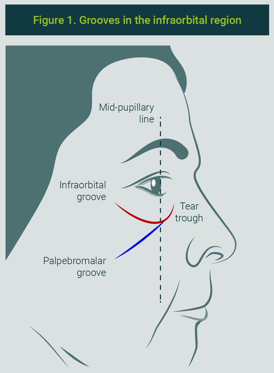
The palpebromalar groove refers to the deep groove that extends approximately 2 cm downward and laterally from the inner canthus of the eye and is commonly seen at the junction of the eyelid and cheek. It comprises the medial third of the nasojugal groove and continues inferolaterally, which in some individuals may be seen visually as the midcheek groove.4,5
During the aging process, skin physiology changes and eventually diminish metabolic processes and normal cellular activities; a process called chronoaging.
One of the first phenomena of skin aging are skin laxity and fine rhytides, which begin at approximately 35 years of age, although they may not become evident until later.The main exogenous factor is photodamage.
Laxity is strictly related to a reduction in collagen and elastin production. It is associated with alterations in the extracellular matrix, particularly with a decrease in the concentration of hyaluronic acid (HA).6
The use of stabilized hyaluronic acid hybrid complexes (Profhilo®, IBSA, Lugano, Switzerland) has gained relevance in aesthetic medicine due to their ability to address skin laxity and optimize skin quality through a process of cellular bioremodeling. This mechanism is based on the interaction between keratinocytes and fibroblasts, which favors the synthesis and organization of extracellular matrix proteins.
Those effects are mediated by the activation of the CD44 receptor, induced by specific molecular weights of HA. Likewise, a significant increase in the production of type I and IV collagen, as well as elastin, has been observed in treated areas, contributing to dermal regeneration.7
The administration protocol for stabilized hyaluronic acid hybrid complexes (HCC-HA) follows a standardized regimen that includes two applications at a 30-day interval.
In each session, 64 mg of non-cross-linked HA are administered, composed of 50% (32 mg) low-molecular-weight HA (80-100 kDa), and 50% (32 mg) high-molecular-weight HA (1100-1400 kDa).
Application is peablrformed by means of 10 intradermal boluses of 0.2 mL each, using a prefilled syringe containing 2 mL of product.
In the case of the face, the injection points have been standardized into five specific points per hemiface, known as Bioesthetic Points (BAP).8
Aliaxin® (IBSA) is a line of dermal fillers based on ultrapure HA, manufactured through a patented biofermentation process using Streptococcus zooepidemicus strains. Its low percentage of cross-linking (<10%) and the minimal level of BDDE residues (<0.1 ppm) allow more effective integration into tissues, reducing trauma and structural alterations compared with other fillers.9
The use of HA fillers for the management of aging of the periocular region has increased as an alternative to surgical procedures with minimal recovery time, low complication rates, and although HA is biodegradable and has limited immunogenic potential.
Nevertheless, fillers may migrate and persist longer than expected and produce edema given the difficult lymphatic drainage of the area.
For this reason, it is crucial to use high-purity HA and fillers with a high cohesivity index to prevent migration and improve tissue integration. Furthermore, a thorough knowledge of facial anatomy and safe injection techniques is essential.10
We hypothesized that the combined use of HA hybrid complexes and cross-linked cohesive HA would produce a synergistic improvement in the infraorbital area, addressing both skin quality through bioremodeling and structural volume loss through mechanical support.
Objective
To present a case series of infraorbital aging with fine rhytides in the lower eyelid, dark circles or displacement of the infraorbital fat pocket in order to demonstrate the efficacy and safety of an intervention procedure with ultrapure and cohesive HA in that specific area of the face in a prospective, non-comparative study.
Justification
Periorbital aging is one of the primary aesthetic concerns because of the central role of the eyes in facial expression and gaze; therefore, these alterations and signs of aging impact on the patients’quality of life. Treatment of dark circles requires detailed anatomical knowledge because many factors may contribute to their appearance.
Eyelid laxity is a disorder that can often go unnoticed, but may contribute to ocular surface pathology and worsen dry eye symptoms because blinking in these patients is less effective.
Genetics contributes to the presence of eyelid bags, but aging plays an important role due to displacement of the infraorbital fat pocket in the setting of bone resorption and ligamentous laxity of the periorbital area.
In this prospective, non-comparative case series, eleven patients (ten women and one man), who attended various private clinics seeking improvement in their infraorbital aging condition (from August to December 2025), were included. All patients signed an informed consent form. The mean age was 45 years.
Inclusion criteria
Patients with dark circles, fine infraorbital rhytides, and malar loss.
Exclusion criteria
Patients with autoimmune diseases, active lesions or infections in the treatment area, keloid scarring, uncontrolled diabetes or hypertension, pregnant women, or those who refuse to sign an informed consent form that includes permission to use their clinical photographs for academic purposes.
Evaluation
For patient assessment, the degree of dark circles was evaluated according to the Hirmand classification, which is a scale used to evaluate tear trough deformity and infraorbital depression. It is divided into three grades based on the severity of tissue atrophy and localized depression:
- Grade 1 (Mild). Slight depression in the tear trough without significant changes in malar fat volume. The skin and the underlying structure still retain good firmness and support. It is generally more evident with lateral lighting. It responds well to treatments with low-cross-linked HA.
- Grade 2 (Moderate). The tear trough depression is more evident and extends laterally toward the malar region. There may be some degree of skin thinning and loss of malar fat volume. More pronounced infraorbital shadows may be noticeable, giving an appearance of fatigue.
- Grade 3 (Severe). Deep tear trough depression with marked loss of volume in the underlying fat. Tissue flaccidity and greater visibility of the transition between the lower eyelid and the cheek. In some cases, protrusion of the infraorbital fat pads is observed.11
Skin condition (fine rhytides and laxity) was assessed according to the modifiedFitzpatrick-Goldman scale12(Table 1) and the Leal Silva scale.
| Table 1. Fitzpatrick - Goldman scale of de rhytides and grade of elastosis to assess the condition of the infraorbital skin | |||
|---|---|---|---|
| Type | Rhytides | Grade | Grade of elastosis |
| 1 | Fine rhytides | 1-3 | Mild (slight textural changes with subtle lines) |
| 2 | Fine to moderate rhytides, moderate number of lines | 4-6 | Moderate (evident elastosis: individual yellowish translucent papules, visible under direct light and dyschromia) |
| 3 | Fine to deep rhytides, numerous lines with or without numerous skin folds | 7-9 | Severe (multipapular elastosis and confluent, yellowish - pale, approaching to or consistent with cutis rhomboidalis) |
The Facial Laxity Rating Scale (FLRS), or Leal Silva scale, is a tool designed to evaluate laxity of the facial skin and soft tissues. It classifies laxity into 10 grades: Grade 0 represents absence of laxity, and grades 1 to 9 indicate a progressive degree of laxity. It also evaluates four distinctive signs in four facial regions13:
- Upper face: eyelid folds
- Midface: nasojugal folds
- Lower face: jowls
- Neck: neck profile
A Global Aesthetic Improvement Scale (GAIS) form was also included; the patients and the treating physicians answered it to treatment satisfaction. Descriptive statistical analysis was used to report the results.
At each visit, patients underwent directed questioning and physical examination of the treated area to actively assess for adverse events, including erythema, edema, ecchymosis, nodularity, or signs of vascular compromise.
Study Design
Three visits were conducted. At the first one (day 0), HCC-HA and Aliaxin GP® were applied, and the following scales were performed:
- Hirmand to assess the degree of infraorbital aging;
- the modified Fitzpatrick-Goldman to assess the condition of the infraorbital skin;
- Leal Silva to assess facial tissue flaccidity;
- and the Likert to grade pain associated with the procedure.
At the second visit (day 30), the second dose of HCC-HA was applied and the Hirmand, modified Fitzpatrick-Goldman, and Leal Silva scales were performed, as well as the Likert scale and a patient GAIS questionnaire.
At the third visit (day 60), no procedure was performed, and the assessments from the second visit were repeated (except for the pain scale), plus a GAIS assessment by the treating physician.
Procedure and Products Used
Aliaxin GP®
Due to its rheology (with a G´ of 95, high cohesivity, a tan delta of 0.23, and a G´ of 0.16), it improves volume in the periocular area, which requires materials with high tissue adaptation in order to avoid undercorrection. Thanks to its cohesivity, gel migration is prevented and greater projection of the material is achieved due to the molecular weights of the HA.
Profhilo®
The eyelid skin is very thin (less than 1mm in thickness), making it especially vulnerable to photodamage and aging. Thus, based on the rheology and the naturally hybrid complex (NAHYCO) technology of this product —which forms hybrid complexes with multilevel action—, it provides hydration to the eyelid skin via its low-molecular-weight chains.
High-molecular-weight HA provides structural support and improves the extracellular matrix through increased tension, preserving the periorbital fat and the rest of the fat pads that interact dynamically with the major support ligaments of the midface.
Application Technique
Patients were evaluated in the seated position to objectively assess the displacement of periorbicular tissues. The depression zone was marked from the inner canthus to the mid-pupillary line, defining the start and end points of the dark-circle depression.
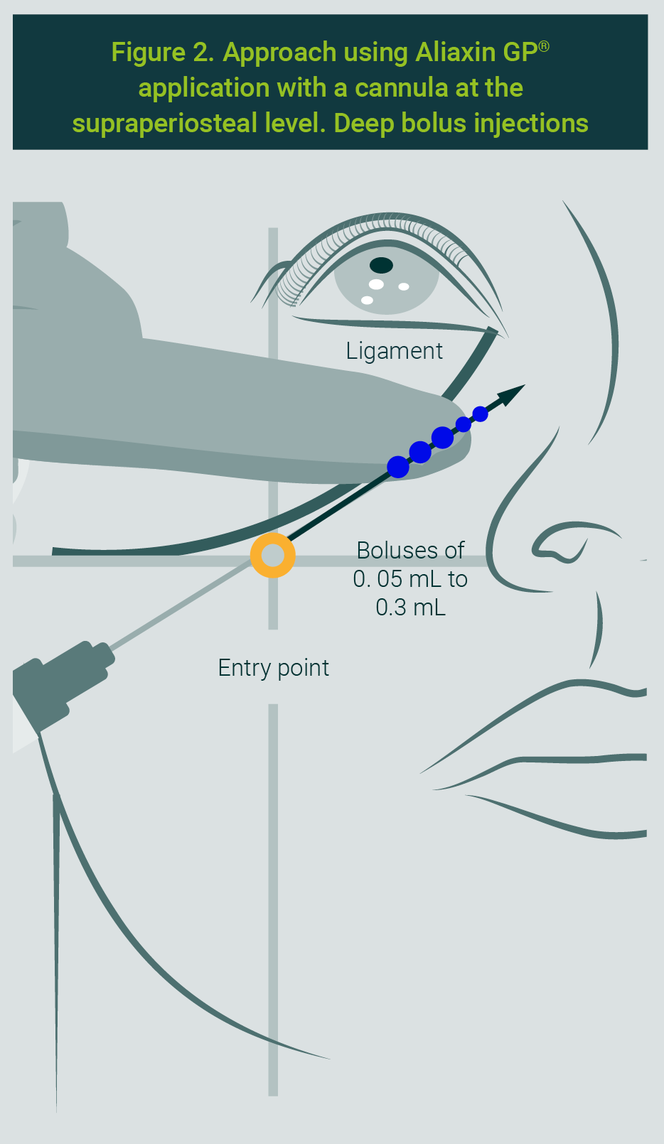
The HA filler was applied with a 22-G or 23-G×50 mm cannula. The entry point was established at the intersection of two lines: one extending from the nasal ala to the earlobe, and the other from the lateral canthus to the jowl. The cannula was introduced in the supraperiosteal plane, applying boluses followed by retroinjection between the boluses, following the trajectory beneath the tear trough ligament and the nasojugal groove.
Boluses of 0.05 to 0.3 mL are recommended to project the tissue according to the patient’s volume replacement needs. Smaller to larger amounts were applied during retroinjection, with the maximum volume targeted at the level of the mid-pupillary line). To ensure precision in application, elevating the cannula is recommended to project the tissue and identify its tip using the hand contralateral to the injecting hand (Figure 2).
For the HCC-HA application, the following marking was performed (Figure 3), derived from the BAP technique:
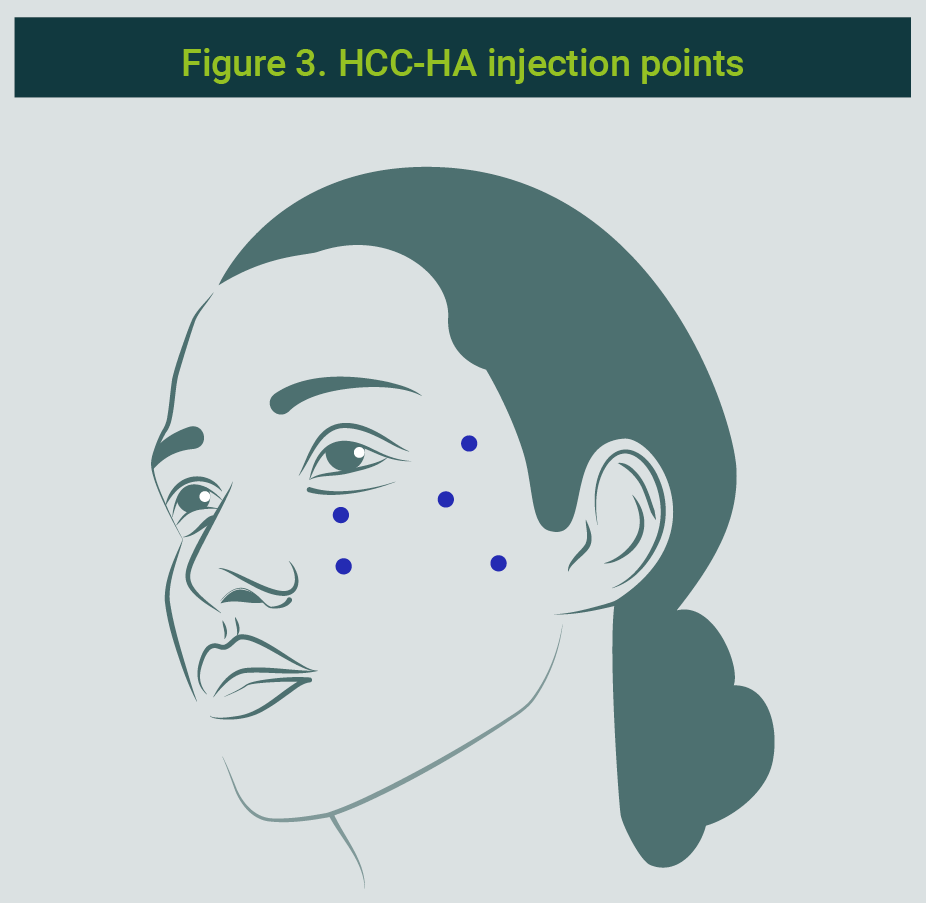
- 0.2 mL at the level of the lateral ocular canthus over the zygomatictemporal junction, aiming to provide multilevel tissue tone to the temporal area —encompassing the lateral orbital fat pads and the lateral temporal region—, and achieving tension by means of the orbicular retaining ligament, the zygomaticocutaneous ligament, the inferior temporal septum, and the SMAS.
- 0.2 mL at 45 degrees from the lateral ocular canthus over the zygomatic bone, aiming to provide firmness to the tissues comprising the main support area of the middle and lower thirds of the face. This preserves the superficial and deep fat pads, such as the deep medial cheek fat, the sub-orbicularis oculi fat (SOOF), and the retro-orbicularis oculi fat (ROOF).
- 0.2 mL on the mid-pupillary line below the orbital rim to preserve fat of superficial and deep fat pads (such as the deep and superficial medial cheek fat, medial SOOF, and medial ROOF), which directly participate in the tension of the zygomatic-cutaneous ligaments.
- 0.2 mL at the intersection of the mid-pupillary line with the line extending from the nasal ala to the tragus, providing support to the infraorbital fatty tissue and the nasolabial fatty tissue.
- 0.2mL at 5cm at the level of the tragus below the zygomaticotemporal suture, aimed at preserving the fat beneath the zygomaticocutaneous ligament, improving tissue tone within the SMAS, and enhancing the tension of the superior portion of the masseteric ligament.
No adverse events were reported in the patients in this study.
Patient satisfaction, evaluated by the GAIS scale, was rated as very positive in 82% of the cases and positive in 18%. The physician’s GAIS rating at the end of treatment showed an 82% very positive and 18% positive.
In the modified Fitzpatrick-Goldman grading (to measure elastosis):
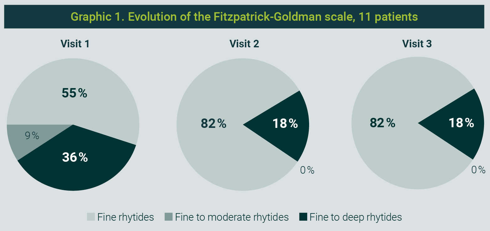
- An initial grading of 36% of patients with grade 3 (severe elastosis) and 9% with grade 2 (moderate elastosis) was obtained; the rest of the patients initially had a grade 1 score (mild elastosis).
- At the second visit, 18% had a grade 3 score and the rest (82%) had grade 1. These scores were maintained at visit 3 (Graphic 1).
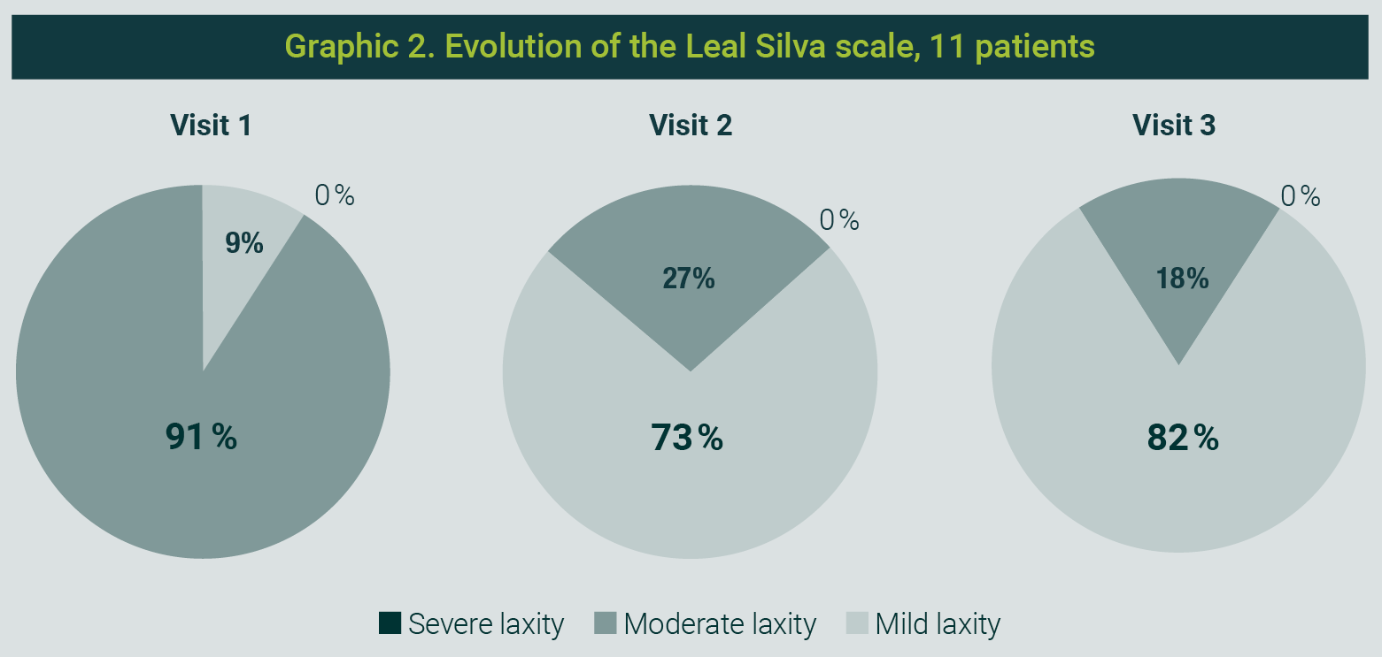
According to the Leal Silva scale for facial tissue laxity:
- At the initial visit, 91% of patients presented with moderate laxity and 9% with mild laxity.
- At the second visit, 27% showed moderate laxity and 73% mild laxity.
- By visit 3, moderate laxity had decreased to 18% and mild laxity was 82% (Graphic 2).
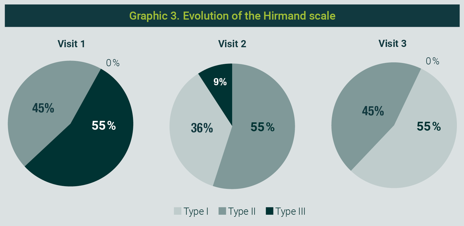
The Hirmand scale, which grades the condition of the infraorbital region, showed:
- At visit 1, that 45% of the patientes were type II (moderate) and 55% were type III (severe).
- At visit 2, 9% were type III, 55% were type II, and 36% were type I (mild).
- At the third visit, 45% were graded as type II and 55% as type I (Graphic 3).
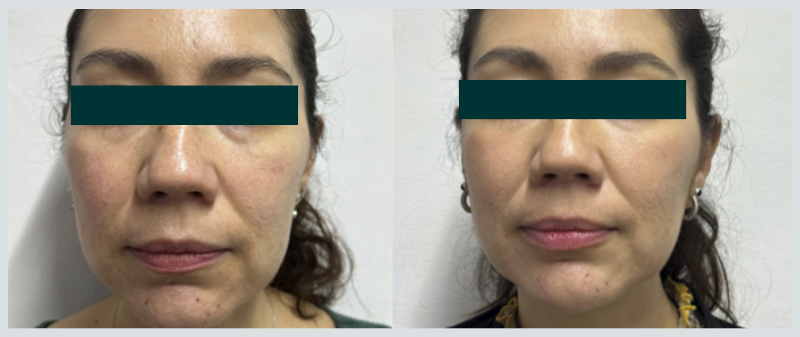
Photograph 1 shows the patient pre- and post-procedure.
The findings of this study support the efficacy and safety of the combined approach using ultrapure cross-linked hyaluronic acid and HCC-HA for the treatment of infraorbital aging.
The improvements observed on the three scales used (Hirmand, modified Fitzpatrick-Goldman, and Leal Silva) reflect not only an improvement in the appearance of dark circles and tissue laxity, but also in the overall quality of the periorbital skin.
This multilevel approach enables action across different tissue layers, with HCC-HA acting as a tissue biomodulator and the ultrapure HA filler providing structural restoration through its high cohesivity and projection capacity. This synergistic effect appears to offer an effective alternative to more invasive procedures, with a high safety profile and no adverse events reported.
The level of satisfaction among patients and treating physicians, assessed through the GAIS scale, was notably high. This suggests that the treatment not only meets objective clinical criteria but also exerts a positive impact on the patient’s aesthetic perception.
Although the sample size is limited and the study was non-comparative, the promising results pave the way for larger, controlled studies that validate this technique as a standard therapeutic option in the management of infraorbital aging.
Combined treatment with high-cohesivity ultrapure hyaluronic acid (Aliaxin GP®) and HCC-HA in the infraorbital area proves to be a safe and effective strategy for improving laxity, fine rhytides, and hollowing of the infraorbital groove.
The results of this study suggest that this approach can improve both deep tissue structure and superficial skin quality, with no adverse events and high levels of satisfaction among physicians and patients. Future studies with larger sample sizes and a control groups are required to validate and standardize this technique.
The authors would like to express their sincere gratitude to Dr. Alfredo Salmon for his valuable support and contribution to this study.
The authors declare they are speakers for the Aliaxin brand. There is no other conflic of interest.
| 1. | Iera M. Treatment of infraorbital skin depressions using the hyaluronic acid filler VYC-15L based on the MD codes approach: A retrospective analysis. J Clin Exp Dermatol Res. 2020;1(4):526. |
| 2. | woodward J, Cox SE, Kato K, Urdiales-Galvez F, Boyd C, Ashourian N. Infraorbital hollow rejuvenation: Considerations, complications, and the contributions of midface volumization. Aesthet Surg J Open Forum. 2023;5:ojad016. |
| 3. | Narurkar V, Shamban A, Sissins P, Stonehouse A, Gallagher C. Facial treatment preferences in aesthetically aware women. Dermatol Surg. 2015;41(suppl 1):S153-S160). |
| 4. | Sadick NS, Bosniak SL, Cantisano-Zilkha M, Glavas IP, Roy D. Definition of the tear trough and the tear trough rating scale. J Cosmet Dermatol. 2007;6(4):218-222. |
| 5. | Niforos F, Acquilla R, Ogilvie P, Safa M, Signorini M, Creutz L, et al. A prospective, open-label study of hyaluronic acid-based filler with lidocaine (VYC-15L) treatment for the correction of infraorbital skin depressions. Dermatol Surg. 2017;43(10):1271-1280. |
| 6. | Uitto J. The role of elastin and collagen in cutaneous aging: Intrinsic aging versus photoexposure. J Drugs Dermatol. 2008;7(2 Suppl):s12-6. |
| 7. | Stellavato A, Corsuto L, D’Agostino A, La Gatta A, Diana P, Bernini P, et al. Hyaluronan hybrid cooperative complexes as a novel frontier for cellular bioprocesses re-activation. PLoS One. 2016;11(10):e0163510. |
| 8. | Rodríguez Abascal M, Saldaña Fernández M. Bio-remodelación facial mediante inyección intradérmica de un complejo híbrido estabilizado de ácido hialurónico de alto y bajo peso molecular: estudio prospectivo en 30 pacientes. Eur Aesthetic Plast Surg J. 2015;5:124-131. [Consultado el 14 de agosto, 2026]. Disponible en URL: https://aecep.es/wp-content/uploads/2018/04/revista_aecep_n22_2015.pdf |
| 9. | La Gatta A, De Rosa M, Frezza MA, Catalano C, Meloni M, Schiraldi C. Biophysical and biological characterization of a new line of hyaluronan-based dermal fillers: A scientific rationale to specific clinical indications. Mater Sci Eng C. Mater Biol Appl. 2016;68:565-72. |
| 10. | Murthy R, Roos JCP, Goldberg RA. Periocular hyaluronic acid fillers: Applications, implications, complications. Curr Opin Ophthalmol. 2019;30(5):395-400. |
| 11. | Hirmand H. Anatomy and nonsurgical correction of the tear trough deformity. Plast Reconstr Surg. 2010;125(2):699-708. |
| 12. | Fitzpatrick RE, Goldman MP, Satur NM, Tope WD. Pulsed carbon dioxide laser resurfacing of photo-aged facial skin. Arch Dermatol. 1996;132(4):395-402 |
| 13. | Leal Silva HG. Facial laxity rating scale validation study. Dermatol Surg. 2016;42(12):1370-1379. |
All Rights Reserved® 2019
Latin American Journal of Clinical Sciences and Medical Technology,Continuous Publication • Responsible Editor: Gilberto Castañeda Hernández • Reservation of Exclusive Use Rights: 04-2019-062013242000-203; ISSN: 2683-2291; both granted by Instituto Nacional del Derecho de Autor. • Responsible for the last update of this issue, Web Master Hunahpú Velázquez Martínez.
Calle Profesor Miguel Serrano #8, Col. Del Valle, Alcaldía Benito Juárez, CP 03100, Ciudad de México, México. Telephone number: 55 5405 1396 • Last modified on August 28th, 2026.
Privacy Policy
Latin American Journal of Clinical Sciences
Latin American Journal of Clinical Sciences (Lat Am J Clin Sci Med Technol) is committed to protecting the privacy and personal data of authors, editors, readers, and other users who interact with its editorial and digital platforms. This privacy policy describes how we collect, use, store, and protect such information.
1. Data controller
Lat Am J Clin Sci Med Technol is responsible for the processing of personal data collected through its website, manuscript submission system, and other official publishing channels.
2. Personal data we collect
Lat Am J Clin Sci Med Technol may collect and use internally (without sharing with third parties) the following personal data:
- Full name
- Institutional affiliation
- ORCID (when the author provided)
- Professional and academic information
- Data associated with manuscript submission, peer review, and editorial processes
- IP address, cookies, and website navigation data
- Administrative information required for scientific publication
No sensitive personal data is deliberately collected.
3. Purpose of data use
Personal data is used exclusively for the following purposes:
- Management of the editorial process (submission, peer review, editorial decisions, and publication)
- Communication between authors, reviewers, and editors
- Indexing, dissemination, and academic archiving of published articles
- Compliance with ethical, legal, and regulatory standards
- Improving website performance and security
- Production of internal statistics on use and editorial performance
4. Legal basis for data use
The processing of personal data is based on:
- The user's explicit consent
- The need to execute the editorial and contractual process
- The legitimate interest of Lat Am J Clin Sci Med Technol in the dissemination of scientific knowledge
- Compliance with applicable legal and ethical obligations
5. Confidentiality and peer review
Lat Am J Clin Sci Med Technol guarantees confidentiality of submitted manuscripts and the peer review process. The data of authors and reviewers will not be disclosed outside the editorial scope, except when strictly necessary for scientific publication or by legal requirement.
6. Data sharing
Some data may be shared only with:
- Editorial management platforms
- Indexing and registration services (e.g., Crossref, PubMed, DOAJ)
- Technology providers needed for website operation
- Competent authorities, where there is a legal obligation
Under no circumstances will the data be sold or used for commercial purposes unrelated to publishing.
7. Data retention
The personal data will be kept:
- For the time required for editorial and publication management
- As long as the article remains published as part of the academic archive
- As required by applicable legal or regulatory obligations
8. Information security
Lat Am J Clin Sci Med Technol implements reasonable technical and organisational measures to protect personal data against unauthorised access, loss, alteration or improper disclosure.
9. Rights of the owners of the information
Users are entitled to:
- Access to their personal data
- Request correction of incorrect information
- Request the deletion of their data, when legally possible
- Object to the processing of their data
- Request the limitation or portability of data
Requests may be submitted through the official channels of Lat Am J Clin Sci Med Technol.
10. Use of cookies
The website of Lat Am J Clin Sci Med Technol may use cookies and similar technologies to improve user experience, analyze traffic and optimize the functioning of the portal. Use of the site implies acceptance of these cookies.
11. Changes to the privacy policy
Lat Am J Clin Sci Med Technol reserves the right to update this Privacy Policy at any time. The modifications will be published on the official website and will take effect upon their publication.
12. Privacy policy acceptance
Submission of manuscripts, registration as a user or use of the website implies express acceptance of this Privacy Policy.